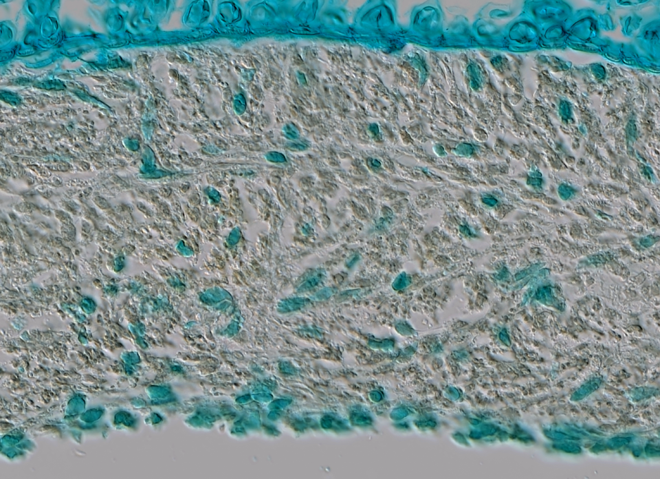
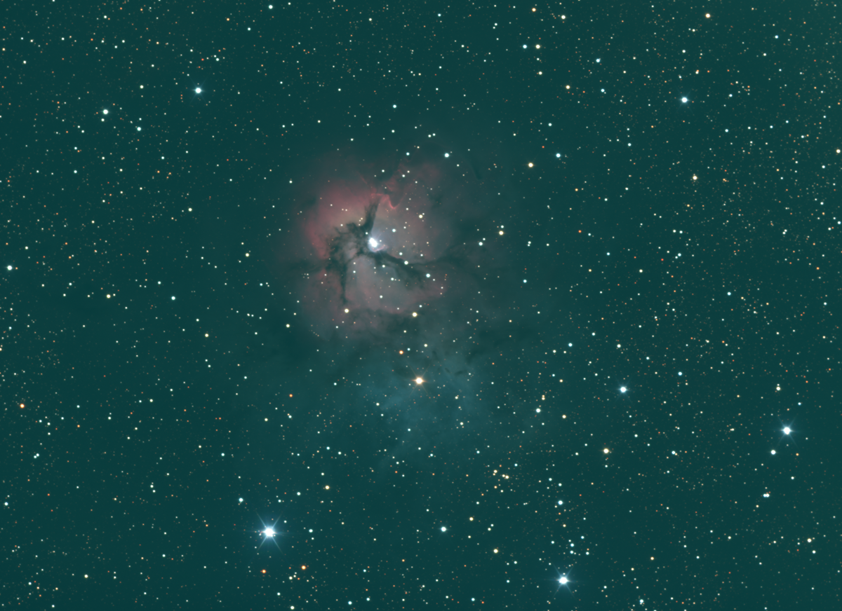
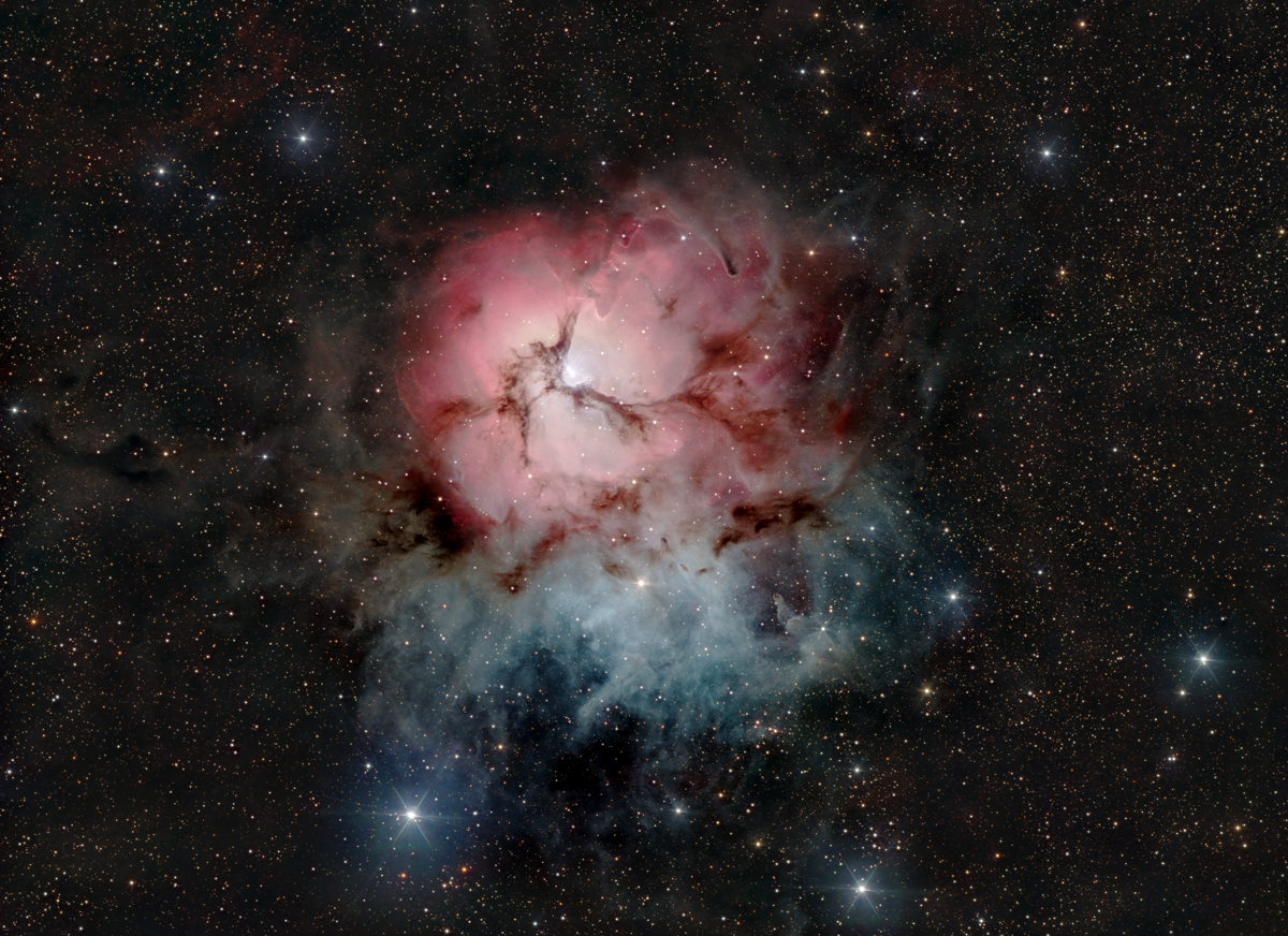
The two images below are of the Trifid Nebula. It shows the raw image vs deconvolution+denoising+histogram stretch.
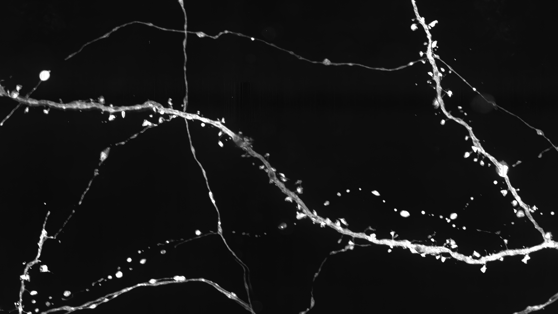
Two images below show a spinning disk confocal microscope image of dendritic spines on a neuron. The image shows vertical striping from the CMOS sensor. The comparison image shows the same image after destriping.


The images below show the raw image and then following denoising and deconvolution.
The images below show denoising. Shot noise is removed but colour noise remains. It is removed further down.
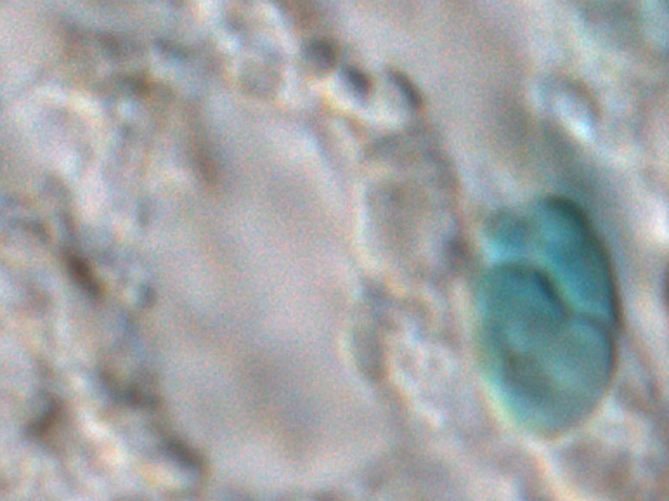
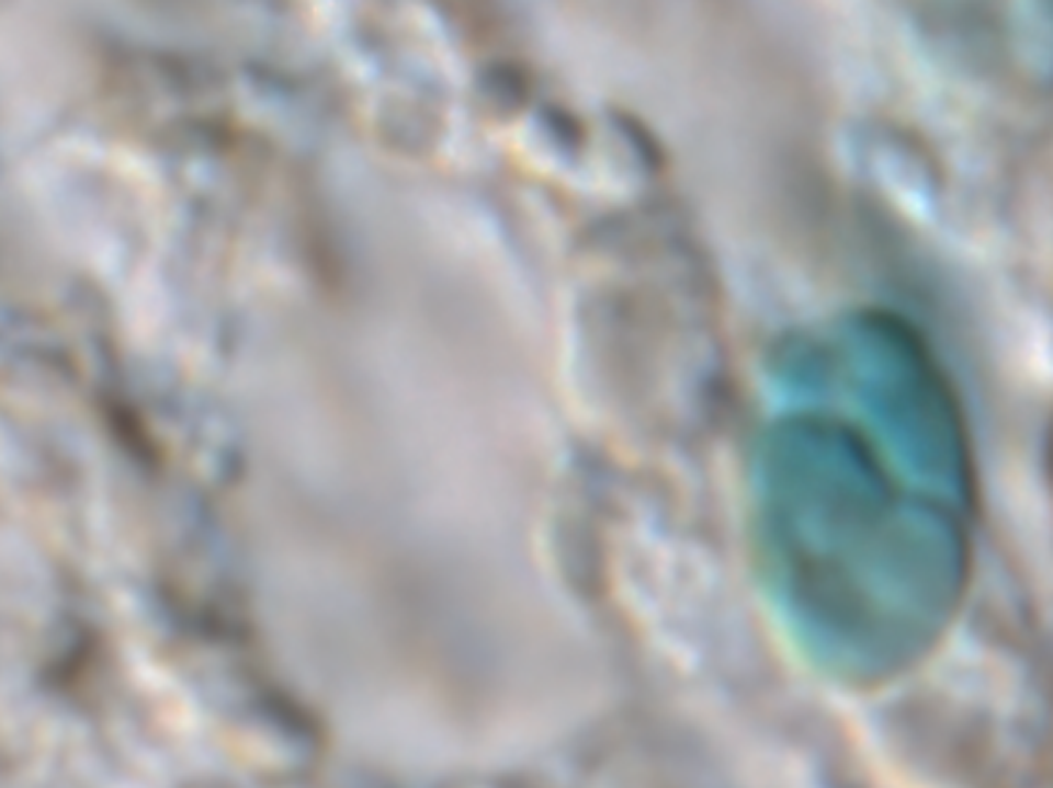
Raw Image
Denoised
Below shows a Denoised vs Denoised+Deconvolved. Two iterations of Regularized van Cittert deconvolution is used.
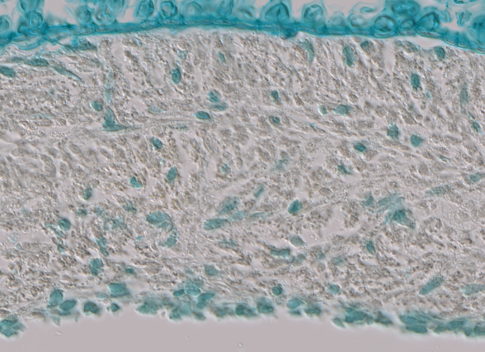
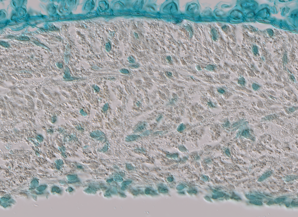
Denoised
Denoised+Deconvolved
Below shows a Den+Decon vs Den+Decon+ColourDenoised. The image is colour-denoised and white balanced.
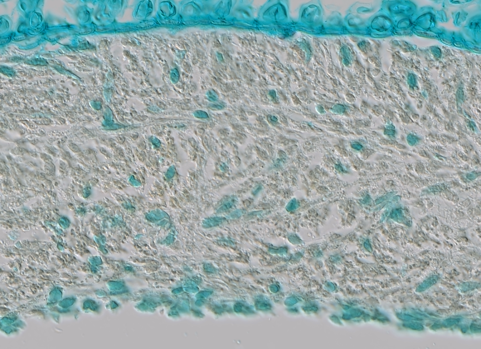
Below shows a DDCD vs DDCD + Levels pair. A histogram stretch is used to remap the black level.